 Contact
 Pharmacie
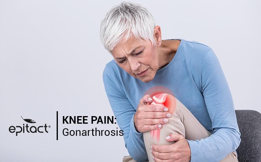
You have knee pain? Your joint is making you suffer? Your knee may be affected by osteoarthritis. According to the World Health Organization, the osteoarthritis is defined as “the result of mechanical and biological phenomenon that destabilise the balance between the synthesis and the destruction of the cartilage and the sub-chondral bone…”. In other words, osteoarthritis is a destruction of the cartilage inside a joint that results of a multifactorial phenomenon. This condition is disabling, chronic, painful and economically expensive.
Gonathrosis is the knee arthritis. Extremely common, this condition is often related to ageing. However, it can affect younger people. This can be linked to a trauma (fracture, ligaments rupture, menisci tearing) or to a sport or work overuse.
The different types of gonarthrosis
The knee joint is made of the femur (thighbone) (at the top) sitting on the tibia (on the bottom) and of the patella on the front. These three bone elements are linked together by passive (e.g.: ligaments, capsule, meniscus…) and active (e.g.: tendon) support structures. So, inside the joint, the femur is articulated through its condyles with the tibial plateau. Between the condyles, there is a groove named trochlea where the patella (kneecap) hinges on. Because of that, there are three areas at risk concerning gonarthrosis:
- The tibiofemoral arthrosis of the internal and/or external compartment, i.e. located between the internal and/or external condyle of the femur and the internal and/or external part of the tibial plateau. That represents 50% of gonarthrosis cases;
- The patellofemoral arthrosis between the trochlea and the patella, that represents around 35% of gonathrosis cases;
- Gonarthrosis combining the two previous represents around 15% of the cases.
The X-ray signs
In order to rule on the advance and the stage of the osteoarthritis, an X-ray exam is needed in addition to a clinical exam. The clinical exam by a general practitioner is essential in order to make a differential diagnosis, which will be then validated by an X-ray. The latest is made standing up, loaded and knee bend on a 30° angle (schuss). We can define 4 stages:
- narrowing of less than 50% of the joint space (stage 1);
- narrowing of more than 50% of the joint space (stage 2);
- a total loss of joint space (stage 3);
- a total destruction of the cartilage between two bones that are directly in contact (stage 4).
The aggravating factors
According to an American study from 2013, 8.5 million UK adults are estimated to have clinical osteoarthritis defined on the basis of symptoms and physical findings. The osteoarthritis before the age of 45 affect only 3% of the people. However, the older people get, the higher the risk is: around 80% of the 80 years-old people are affected. Women are twice as likely to be affected. This condition also concerns more people with high Body Mass Index and that present the factors associated with co-morbidities.
So, across several studies, different factors have been identified as being aggravating the appearance of gonathrosis.
- age;
- gender;
- weight;
- anatomical axial misalignment (genu valgum or varum, coxa vara or valga);
- postural axial misalignment (pronation or supination of the foot);
- important muscle imbalances;
- past traumas (fracture or ligament rupture);
- sport and/or professional overwork;
- inflammatory or metabolic disease.
The treatments
An American study conducted in 2007 emphasizes that by the year 2030, there will be an increase of 673% of surgery for a knee prosthesis. Surgery must however be a matter of last resort. Numerous treatments exist before this drastic method. Here some treatments that tend to relieve pain related to the gonathrosis:
- physiotherapy sessions;
- wearing orthopaedic soles;
- wearing a knee brace;
- drug treatment;
- nutritional follow-up;
- corticoids or hyaluronic acid injections;
- surgery, either for a knee replacement or for a correction of the bone misalignment osteotomy.
The EPITACT® solution
To relieve knee pain, EPITACT® has developed a suited knee brace: PHYSIOstrap™ MEDICAL*. It is made up of several elements that makes it unique on the market.
One of these elements is the EPITHELIUMFLEX® simulated tendon. This patented technology associates a high-performance technical fabric and silicone. Its specific pattern allows during the flexing movements to guide the kneecap in the femoral trochlea. As 'guided', the kneecap won’t be able to dislocate.
Furthermore, the technical fabrics of this knee brace offer a compression and a stability feeling. Developed to offer the right compression, they improve the comfort without impairing the movement. Indeed, at the back of the knee (in the popliteal fossa), the fabric has been specifically conceived to not create discomfort or irritation when it is worn, even after a long period of sitting position.
This knee brace is also made of two silicone elastic strips. Located on the thigh area, these strips ensure a good support all day long. Lastly, facing the patella, an extra-thin and flexible fabric surrounded by a patented silicone tendon that avoid all pressure on it. And the all device weigh only 40 grams! It provides you joint stability and comfort while being worn with discretion under your clothes.
*This product is a class I medical device that bears the CE marking under this regulation. Carefully read the instructions before use. Manufacturer: Millet Innovation. 09/2020
For more details about this general and simplified approach, here are further sources:
The epidemiology and impact of pain in osteoarthritis, T. Neogi, Clinical Epidemiology Research and Training Unit, Boston University School of Medicine, Boston, MA, USA, Osteoarthritis and Cartilage 21 (2013)
Guillemin F, Rat AC, Roux CH, Fautrel B, Mazières B, Chevalier X et al.; KHOALA cohort study. The KHOALA cohort of knee and hip osteoarthritis in France. Joint Bone Spine 2012; 79:597-603.
Kurtz S, Ong K, Lau E, Mowat F, Halpern M. Projections of primary and revision hip and knee arthroplasty in the United States from 2005 to 2030. J Bone JointSurg Am 2007;89:780-5
Professeur Dominique SARAGAGLIA, Les indications chirurgicales dans la gonarthrose, Corpus Médical – Faculté de Médecine de Grenoble, Mars 2003
Richette P. Généralités sur l’arthrose : épidémiologie et facteurs de risque. EMC - Appareil locomoteur. janv 2008; 3(4):1‑5.